

 By
Decoding Cellular Communication: How AI is Revolutionizing Our Understanding of Cell Development
Decoding Embryonic Development: Breakthrough by MIT Researchers
Embryonic cells, originally just stem cells, each take on individual roles as they evolve. They begin to differentiate, transforming into various types of cells, such as brain, liver, or muscle cells. These changes lead to the construction of three-dimensional structures, like tissues and organs. Each cell’s destiny, including its particular specialization, is orchestrated by the activation or silencing of specific genes. These patterns of gene activity then shape the cell’s structure and function, enabling it to perform a unique role in the body.
However, this process is far from solitary. Cells are continuously communicating, exchanging chemical signals with their neighbors. This helps them take stock of their surroundings, realize their stage in development, and prepare for their ultimate fate. To do this, messages are sent over signaling pathways, a series of steps that convert external signals into changes in gene activity within the cell.
A New Approach to Mapping signaling Pathways
For scientists, unraveling this intricate language of cells is key to understanding the development of tissues, diseases, and how to correct malfunctions. But this deciphering task has traditionally been challenging, as it was commonly believed that the effects of signaling pathways differed vastly across cell types. This supposed variability deemed it necessary to create separate maps for each cell type, which is a laborious and intricate task.
However, recent breakthroughs by researchers from MIT indicate this process may not be as arduous as we previously believed. A study led by Pulin Li and graduate student Nicholas Hutchins discovered that each signaling pathway leaves a unique ‘fingerprint’, a distinctive pattern of gene activity that offers clues to the signals a cell has encountered. Remarkably, these fingerprints are consistent across different cell types for the same signaling pathway. Thanks to this consistency, scientists can now use these pathway-specific fingerprints to reconstruct signalling pathways across multiple cell types, eliminating the need for individual mappings.
IRIS: Harnessing AI for Cellular Studies
This groundbreaking discovery was powered by a machine learning model named IRIS. IRIS is sophisticated enough to detect different fingerprints of signaling pathways, and determine the signals a cell received throughout various developmental stages, even in unexplored cell types. This technology presents a significant leap over traditional methods, which were reliant on experimenting on every possible pathway in every cell type. It breaks open the possibilities for comprehensive mapping of signaling histories of each cell within a mouse or human embryo on an unprecedented scale.
The researchers’ findings, featured in the esteemed Nature Methods journal, could dramatically speed up the engineering of stem cells for regenerative medicine. It could also lead to advancements in the generation of organoids, miniature, 3D models of real organs created for disease study and drug experimentation. Identifying the signal patterns that prompt a stem cell to become a certain cell type could help researchers in controlling stem cell fate, both in labs and in medical contexts.
IRIS, which is a neural network-based model, is like an AI-driven system developed to recognize patterns in intricate data, much like our brains decode patterns. It looks at a cell’s overall gene activity and estimates the signaling pathways likely active during development. The researchers trained IRIS using an expansive dataset, charting how thousands of human embryonic stem cells responded to combinations of six main signaling pathways at various stages. This training led to the creation of a comprehensive map reflecting how signaling combinations influence cell behavior.
To give a glimpse of IRIS’s real-world application, the team tested it using single cells from mouse embryos during gastrulation, a stage where cells rapidly begin divvying up their roles. IRIS accurately predicted when and where specific signaling pathways activated in cells that were destined to form heart, gut, muscle, and spinal cord tissue. The variation in signaling molecules instructs cells to form the correct structures in their rightful places.
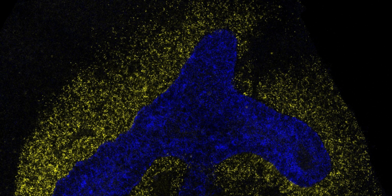
The researchers also used IRIS to pinpoint the necessary signals for forming a cell type crucial for lung development. Following the model’s prediction, an experiment in mouse embryos confirmed that stimulating a specific signaling pathway encouraged lung-specific growth. By identifying the exact combinations of signals that drive lung cell development, researchers can engender lab-grown models of lung tissue with greater reliability.
“IRIS is helping us decode the language cells use to communicate at a much faster rate than traditional experiments could realistically achieve,” says Hutchins. He adds that these models would allow researchers to study diseases like asthma, lung cancer, and pulmonary fibrosis, where lung tissue becomes scarred, often without a known cause. Such models can then be used to test potential therapies, and ultimately help design regenerative treatments that can mend damaged tissue.
Accompanying technological advancements with implementi.ai could aid companies in integrating AI automation into their operations and uncover ways to revolutionize their businesses.
More details on this monumental study can be found on MIT News.


