
AI Tool Offers Unprecedented Insight into Brainstem’s Vital White Matter Pathways
As we delve deeper into the depths of the human mind, the brainstem is a focal point of study. This vital hub controls many of our body’s core functionalities, from sleep and consciousness to breathing and heart rate. Yet, visualizing it with clarity has been a challenge due to the intricate bundles of white matter fibers that power these operations. Traditional imaging systems have struggled to provide detailed visuals of these fibers, keeping clinicians and researchers in the dark on how these areas change response to trauma or disease.
In a collaborative effort, researchers from MIT, Harvard University, and Massachusetts General Hospital have made groundbreaking strides. They have developed an innovative AI-powered tool called BrainStem Bundle Tool (BSBT) to shed some light on this unexplored region of the brain. Their findings were published in the Proceedings of the National Academy of Sciences, and the tool is now publicly accessible on GitHub.
A New Dimension to Brainstem Studies
The BSBT was born out of the doctoral research of Mark Olchanyi, the MIT graduate student who spearheaded the study. He saw the need to understand the organization of the white matter in humans and how it deteriorates in certain disorders. Olchanyi and his team took advantage of diffusion MRI, a specialized imaging method that tracks how water moves through the brain’s white matter, thereby revealing the structure of neural pathways.
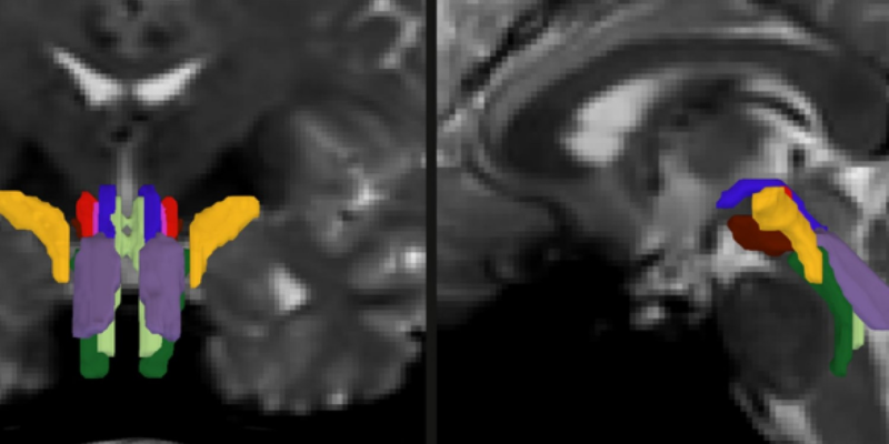
However, imaging the brainstem posed significant challenges due to its small size and its close proximity to pulsating fluids and moving organs. This is where the AI model steps in. It uses a convolutional neural network to analyze diffusion MRI scans, creating a probabilistic map of fiber pathways that descend into the brainstem. It then delves deeper to identify eight specific white matter bundles within the brainstem.
Testing The Tool
For BSBT to become a reliable tool, it had to be put through rigorous testing and validation. Olchanyi trained the system using 30 annotated diffusion MRI scans from the Human Connectome Project. The tool’s accuracy was then cross-verified by comparing its results with data from post-mortem brain dissections and ultra-high-resolution scans. BSBT’s reliability was confirmed when it successfully identified the same bundles in repeated scans of the same individuals.
Once validated, the system was put to use analyzing scans from patients with various neurological conditions, including Alzheimer’s, Parkinson’s, multiple sclerosis (MS) and traumatic brain injury (TBI). The tool proved efficient, distinguishing between patients and healthy controls better than other methods, signifying its potential usage as a diagnostic aid.
One remarkable test case was of a 29-year-old man who had endured a severe brain (TBI) injury and was in a coma for seven months. The tool helped in imaging his brainstem bundles which, fortunately, were not severed despite being displaced. Over time, BSBT tracked the decrease in size of the lesions on the bundles and their gradual return to normal positions, echoing the patient’s recovery.
Looking into the Future of Brain Health
Under the guidance of Professor Emery N. Brown, Olchanyi’s thesis advisor and a co-senior author of the study, they have enhanced our understanding of complex brain functions by gaining access to the crucial brainstem area. BSBT is truly groundbreaking; it represents a significant advancement in neuroscience research and clinical diagnostics. Its open-source availability paves the way for worldwide studies in brain health and disease, making brainstem studies more accessible and in-depth than ever before.
For more details, read the original news release here.


